Written by Microscopehunt Authority |
.
Explore the hidden creatures around you with a microscope! Watch tiny creatures come to life, learn about the parts of a microscope with functions, and take stunning close-up photos.
.

The microscopes are wonderful Instruments that changed our perspective of the microscopic world. They enable scientists, students, and those with inquisitive minds to examine structures and organisms that are too small to see without technology. From the detailed blueprints of cells to the tangled clustering of microorganisms, the ability to see these small intricate details has broadened the frontiers of biology, medicine, and materials science. In this guide, we will explore the different parts of a microscope and what each part does, and hopefully, by the end, we will have an understanding of this essential scientific tool.
Introduction to Microscope: Parts of a Microscope with Functions
In the late 16th century inventors like Zacharias Janssen and Antonie van Leeuwenhoek began crafting lenses to magnify small objects.Microscopes have transformed from basic optical devices into complex instruments which provide deep insights into life’s smallest components. Today, optical, electron, and scanning-probe microscopes find roots in one another, but their traits and capacities are different from one other. To be able to use them accordingly, it is important for anyone interested in using a microscope to understand the different parts of all these variations.
The Basics: Understanding the Essential Parts of a Microscope

A microscope may seem like a complicated piece of equipment at first glance, but it is really all about an arrangement of carefully aligned components working together. If you have a microscope, you can see that the components of the scope are magnifying and illuminating the specimen you look at. Known as the basic parts of a microscope, regardless if you are working with a simple compound microscope or an advanced electron microscope, they won’t differ significantly. Now, let’s dive into these core components and their roles.
Optical Components: Lenses That Magnify Our World
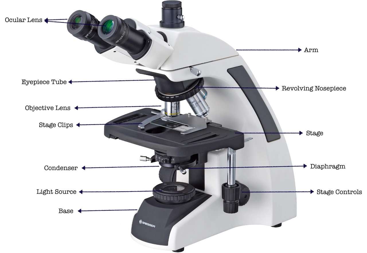
Without a doubt, the most crucial parts of a microscope are its optical components. Two lenses make up this part of the microscope, the eyepiece (also known as the ocular lens) and the objective lenses that work in short to magnify the specimen. The eyepiece is the lens that’s closest to your eye, so it generally comes with a 10x magnification. The objective lenses, which are located on the rotating nosepiece, come in a variety of magnifications, including 4x, 10x, 40x, and 100x.All these lenses amplify their magnifying power cumulatively, letting you see details that would otherwise be far beyond detection.
The clarity and distortion free images that one can expect from these lenses depends on the quality of the lenses themselves. Well-made microscopes consist of precision-ground glass lenses with anti-reflective coatings that reduce distortions and improve resolution. It is the optical devices within the microscope that enables it to unveil the wonders of the microscopic world.
Illumination Systems: Shedding Light on the Micro Universe
To observe specimens under a microscope with clarity and detail, a proper illumination system must be employed. If the specimen is not properly illuminated, even the most powerful lenses would not be useful. Nowadays, most advanced microscopes are equipped with light sources mounted below the stage, such as a built-in LED or halogen bulb. This light is then frosted through a condenser which guarantees that the specimen is evenly lit.
An equally important aspect of the illumination system is the diaphragm, which regulates the intensity of light that hits the specimen. Altering the diaphragm can further improve sharpening and contrast, enabling easier examination of details. Be it transparent cells or opaque materials, the illumination system plays a significant part in focusing the specimen.
The Stage: Where Specimens Come to Life
The stage is the part of the microscope where the slide containing the specimen is kept for observation. It usually is a rectangular slab which is flat and has either clips or mechanical holders to keep the slide stationary. Some types of microscopes have a mechanical stage that enables you to move the slide with knobs, while others have a fixed stage that can only be adjusted manually.
The stage is vital to the microscope since it makes sure that your specimen is safe and focused. With no stable stage, any movement at all would completely alter your view, making it nearly impossible to study in closer detail. So no matter if you are looking at a single cell or even a highly detailed tissue sample, the stage is what forms the basis for all good microscopy.
Focus Mechanisms: Achieving Clarity in Microscopic Views
One of the most important features of microscopy is obtaining a clear and sharp image, and this is where the focus mechanisms are important. Most microscopes come with two focus knobs, the coarse focus knob and the fine focus knob. The coarse focus knob brings the specimen into general focus, while the fine focus knob adds precise tuning for optimal clarity.
For viewing specimens with different magnifications, these focus mechanisms are very important. When switching from low power to high power, the fine focus knob becomes essential for resolving finer details. Achieving the detail that is known for microscopes without these mechanisms would be almost impossible.
The Body Tube: Connecting Vision and Precision
The body tube is the structural element that connects the eyepiece with the objective lenses. This alignment is crucial to ensure that light travels directly and in a straight path from the objective lens to the eyepiece. In some microscopes, the body tube also contains prisms or mirrors to deflect light and improve image quality.
The body tube is also fine-tuned for length and configuration for optimal output. The image quality also will be distorted if this part is misaligned or damaged, which confirms this commonly neglected part is the most important one.
Eyepieces and Objectives: The Gateway to Detailed Observation
The eyepiece and objective lenses are the main elements involved in magnification. The eyepiece (or ocular lens) is what you look through to view the specimen. It usually has a10x magnification power and functions along with the objective lenses to get a higher magnification.
The objective lenses, located on a rotating nosepiece, provide differing degrees of magnification. Rotating the nosepiece allows you to switch between lenses to view the specimen from various scales. Imaging the fine details requires high-quality objectives, which are thus a crucial component of any microscope.
The Role of the Diaphragm: Controlling Light for Optimal Imaging
The diaphragm is a small yet powerful part that manages the amount of light that reaches the specimen. Found beneath the stage, it features an adjustable opening that can be modified to control light intensity. Correctly adjusting the diaphragm is crucial for obtaining the best contrast and clarity, particularly when examining transparent or low-contrast specimens.
By adjusting the diaphragm, you can improve the clarity of fine details and minimize glare, which makes it easier to examine your specimen. This part is especially crucial in brightfield microscopy, where adequate lighting is essential for effective observation.
Mechanical Stage vs. Fixed Stage: Navigating Your Specimen
When discussing stages, microscopes typically feature two main types: mechanical and fixed. A mechanical stage enables precise slide movement through knobs, facilitating the navigation of larger specimens or targeted areas. This design is particularly beneficial in research and educational environments, where accuracy is crucial. Conversely, a fixed stage necessitates manual slide adjustments, which may lack precision but can be adequate for basic observations. Each type has its own benefits, and the decision between them hinges on your particular requirements and uses.
The Importance of the Base: Stability in Microscopy
The base of the microscope serves as its foundation, offering stability and support for all the other components. A robust base is crucial for minimizing vibrations and ensuring that the microscope stays steady while in use. This is especially vital at high magnifications, where even the tiniest movement can interfere with your view.
Beyond providing stability, the base typically contains the light source and power controls, making it a key part of the microscope’s functionality. Without a strong base, the microscope would struggle to produce the precise, high-quality images that users depend on.
Accessories and Add-ons: Enhancing Your Microscopic Experience
The basic parts of a microscope are crucial, but many extras and add-ons can make your microscope work better. These include things like camera attachments to take pictures special lenses for specific uses, and filters to improve contrast and color. Some microscopes also come with programs to analyze images letting you measure and study your samples more . These extras can turn a simple microscope into a strong tool for research, teaching, and hobby exploration. By getting the right add-ons, you can open up new possibilities and step up your microscope work.
Common Challenges: Troubleshooting Microscope Components
Even top-notch microscopes can run into problems now and then. Users often face issues like fuzzy images patchy lighting, and trouble getting things in focus. These snags stem from lenses that aren’t lined up right dirty parts, or a diaphragm that’s not set . To avoid these headaches, it’s smart to clean and take care of your microscope . This helps it work as well as it should. If you keep running into trouble, you might want to check out the manual that came with your microscope or ask an expert for help. By dealing with these problems , you can keep your microscope working well and giving you results you can trust.
FAQ
- What is the most important part of a microscope?
Even though all the parts are of great significance, the optical elements are probably the most important because they decide the quality and magnification of the picture. - How should I clean the microscope lens?
For cleaning lenses you can use a photonic lens cleaning solution and for gentle cleaning of the turret of the lenses you may just try a microfiber towel. The sole objective of this cleaning process should be taking off any dirt or dust particles and at the same time, you must avoid using any harsh chemicals and abrasive materials that can be harmful to the optics. - How to upgrade my microscope with better components?
Definitely, You can either do it yourself or purchase a deluxe model since there are numerous microscopes available on the market and some of them have a possibility to be upgraded by changing or adding new accessories like the objective, the eyepiece, and the illumination system for better performance. - What is the difference between a compound and a stereo microscope?
A compound microscope is used for watching the small, transparent samples under high magnification, whereas a stereo microscope brings a larger scale and is a tool for observing three-dimensional objects.
Conclusion:Parts of a Microscope with Functions
The progress in the field of microscopy is astounding, and it all starts with understanding the parts of a microscope with functions. Each of the parts of a microscope with functions plays a vital role in enabling us to explore the microscopic world. From the lenses to the light sources, every component of the microscope contributes to the overall ability to magnify and illuminate tiny organisms. By understanding the parts of a microscope with functions, we gain a deeper appreciation for how these instruments work to reveal details that are otherwise invisible to the naked eye. Whether you are a scientist, educator, or hobbyist, knowing the parts of a microscope with functions is key to unlocking the wonders of the microscopic world. The parts of a microscope with functions empower us to take stunning images and make groundbreaking discoveries in many fields.
